Review On -"Wheat grass"

*INTRODUCTION
Wheat grass can be traced back in history over 5000 years, to ancient Egypt and perhaps even early Mesopotamian civilizations. It is purported that ancient Egyptians found sacred the young leafy blades of wheat and prized them for their positive effect on their health and vitality. The consumption of wheatgrass in the Western world began in the 1930s as a result of experiments conducted by Charles F. Schnabel in his attempts to popularize the plant1. By 1940, cans of Schnabel's powdered grass were on sale in major drug stores throughout the United States and Canada[1].

Throughout human history, plants have played a key role in treating human diseases. In thousands of years of trials, human found many plants which are good for treating ailments and curing serious health problems like cancer, diabetes, and atherosclerosis. They are a kind of alternative medicine that is inexpensive, and has no side effects. For example: wheatgrass, aloe vera, curcumin, alfalfa, garlic, ginger, German chamomile, grapefruit, green tea. In 2002, the U.S. National Center for Complementary and Alternative Medicine of National Institutes of Health began funding clinical trials about the effectiveness of herbal medicines[2]. Wheatgrass, has been an integral part of Indian culture for thousands of years, and has been known to have remarkable healing properties. Scientifically known as Triticum aestivum, it belongs to Poaceae family. Other plants included in this family are: Agopyron cristatum, Bambusa textilis, Cynodon dactylon, Poa annua, Zea mays, Aristida purpurea etc. There is not much scientific data available on these plants because of a lack of substantial research. Therefore, it is important to study their properties to explore their maximum benefits. Wheatgrass’ culms are simple, hollow or pithy, glabrous, and the leaves are approximately 1.2 m tall, flat, narrow, 20-38 cm long and 1.3 cm broad3. The spikes are long, slender, dorsally compressed and somewhat flattened Phytochemical constituents of wheatgrass include alkaloids, carbohydrates, saponins, gum and mucilages. Its water soluble extractive value is found to be greater than its alcohol soluble extractive value. This is because of the chlorophyll content of wheatgrass, which is about 70% water soluble[3].

Wheatgrass juice is high in vitamin K, which is a blood clotting agent. People taking bloodthinning medications or people with wheat related allergies shouldn't drink wheat grass juicewithout consulting a healthcare professional. Wheat allergies are generally a response to the gluten (a protein) found in the wheat berry[4]. The environment in which wheatgrass grows determines its vitality and is thus sown in late autumn for maximum concentration of the active principles. The nutritional vibrancy of wheatgrass is encouraged by supplementing the soil with rich vegetable compost and seaweed. At the onset of the spring season, the simple sugars produced as a result of photosynthesis, undergo conversion into proteins, carbohydrates and fats, with the aid of the various enzymes and minerals absorbed by the plant via its roots. Due to the
comparatively lower temperatures in the spring, the grass grows slowly enough for this conversion to occur before the critical jointing stage of growth. At jointing, or the reproductive stage of the plant, the nutrients and energy of the plant are redirected to seed formation. Wheatgrass is harvested just prior to this jointing stage, when the tender shoots are at their peak of nutritional potency[5].

The major clinical utility of wheatgrass juice is due to its antioxidant action which is derived from its high content of bioflavonoids like apigenin, quercitin and luteolin. Other compounds present, which make this grass therapeutically effective, are the indole compounds, choline and laetrile (amygdalin). In a study conducted to determine the elemental concentration profile of wheatgrass using instrumental neutron activation analysis, it was found that the concentration ofelements such as K, Na, Ca and Mg increased linearly in the shoots with the growth period whereas the concentrations of the elements namely Zn, Mn and Fe remained constant in shoots after 8th day of plant growth for all three conditions of growth. However, it was observed that theshoot to root concentration ratio in all the conditions increased linearly for K, Na, Ca, Mg and Cl and decreased for Zn, Fe, Mn, and Al with growth period[6].

* CHEMICAL COMPOSITION OF WHEAT GRASS:-
The major chemical constituents that make wheat grass a valuable food are[8-9]:

PROTEINS Essential and dietary non essential amino acids like leucine, iso leucien, threonine, valine, threonine, phenylalanine, tryptophane, metheonine, lysine, arginine aspartic acid, glycein, prolein, glutamic acid, alanine, tyrosine are present in wheat grass.

VITAMINS Wheat grass contains vitamin A, carotene, B-complex, E, C and K.

MINERALS Iron, calcium, phosphorus, megnasium, zinc, copper, sodium, sulfur, boron, molybdenum, iodine are the important minerals present in wheat grass.

CHLOROPHYLL Wheat grass juice is also known as green blood as it contains chlorophyll. It neutralizes infection, heals wound, overcome inflammation, and gets rid of parasitic infection. Blood purification, liver detoxification and colon cleansing are the three important effects of wheat grass on human body [9-10].

ENZYMES Protease, amylase, lipase, cytochrome oxidase, trans hydrogenase, superoxide dismutase enzymes are present in wheat grass.

LIPASE Lipase is a highly effective in the digestion of fats. Enhances the digestion of proteins, starch and fat in the gastrointestinal tract. Without lipase fat stagnates and accumulates in the organs, arteries and capillaries.
·         
       CYTOCHROME OXIDASE
      Major effector in the body’s production of energy. Cytochrome oxidase anchors a chain of enzymes in the mitochondrion; the power plant of the cell enables this by reacting with oxygen to make energy.
·         
       CATALASE
      This enzyme is among the most efficient known. Serves to protect each individual cell from the toxic effect of hydrogen peroxide. Hydrogen peroxide is caused in the body by bacteria.
·        
      MALIC DEHYDROGENASE
       Important enzyme in maintaining the body’s ability to defeat bacteria and other parasitic hosts in the body.
·        
      ABSCISIC ACID
       Anti-cancer agent.
·         
      PROTEASE, AMYLASE
      Important in supplementing the body’s natural digestion of starches, proteins, fats and cellulose. Can help offset the worst aspects of digestive leukocytosis, the immune response to food heated over 118 degrees.

·         BIOFLAVANOIDS
      Apigenin, quercitin, luteonin are found in wheat grass.

* Pharmacological activity of Wheat Graces juice:

5.1 Hemoglobin and Chlorophyll
Wheatgrass is rich in chlorophyll and enzymes. It contains more than 70% chlorophyll (which is an important dietary constituent). The chlorophyll molecule in wheatgrass is almost identical to the hemoglobin in human blood. The only difference is that the central element in chlorophyll is magnesium and in hemoglobin it is iron [11] (Figure 4). The molecular structure of chlorophyll in wheatgrass and hemoglobin in the human body is similar, and because of this wheatgrass is called 'Green Blood' [6]. A 70-83% increase in red blood cells and hemoglobin concentration was noted within 10-16 days of regular administration of chlorophyll derivatives [12]. It was reported that chlorophyll enhanced the formation of blood cells in anemic animals [13]. Chlorophyll is soluble in fat particles, which are absorbed directly into blood via the lymphatic system. In other words, when the ―blood‖ of plants is absorbed in humans it is transformed into human blood, which transports nutrients to every cell of the body. Chlorophyll present in wheatgrass can protect us from carcinogens; it strengthens the cells, detoxifies the liver and blood stream, and chemically neutralizes the polluting elements.

5.2 Wheatgrass in Cancer prevention
Environmental factors play an important role in the multistage process of cancer development, and nutritional intervention has been identified to play a very important role in its prevention. Dietary compounds such as garlic, carotenoids, wheatgrass, etc are important due to their antioxidant properties. These dietary products protect against many diseases because food and degraded products come into direct contact with bowel mucosa, and can influence its physiology and metabolism. Although many dietary compounds have been suggested to contribute to the prevention of cancer, there is a strong likelihood that wheatgrass extract, which contains chlorophyll, an antioxidant, may affect cancer prevention. Additionally, selenium and lactrile present in wheatgrass have anti-cancer properties[8]. Selenium builds a strong immune system, and can decrease the risk of cancer . Wheatgrass contains at least 13 vitamins (several of which are antioxidants) including B12, abscisic acid, superoxide dismutase (SOD), cytochrome oxidase, mucopolysaccharide . SOD converts two superoxide anions into a hydrogen peroxide molecule, which has an extra oxygen molecule to kill cancer cells.
Although most people use wheatgrass as a dietary supplement or as serving of vegetables, some proponents claim that a dietary program commonly called wheatgrass diet can cause cancer to regress and extend lives of people with cancer . The true cause of the cancerous degeneration of cells has been revealed to be from the destruction of a specific respiratory enzyme, cytochrome oxidase . P4D1, a glycoprotein present in wheatgrass, also acts similarly to antioxidants, stimulating the renewal of RNA and DNA. It is alsop thought to protect the body from the attack of cancer cells by making the walls of cancer cells more op12en to attack by white blood cells . So, the use of wheatgrass in terminally ill cancer patients should be encouraged . It was determined that chlorophyll is an active component in wheatgrass extract, which inhibits the metabolic activity of carcinogens . Adjuvant fermented wheatgrass extract (Avemar nutraceutical) improves survival of high-risk skin melanoma patients . Karager et al has concluded that wheatgrass extract inhibits proliferation of 32Dp210 (BCR-ABL fusion gene (+) mouse CML cell line) cells through the induction of apoptosis[12]

5.3 Hepatoprotective role of wheatgrass
Triticum aestivum leaf extract affects liver enzyme activities as well as lipid peroxidation [10]. Jain et al reported the hepatoprotective role of fresh wheatgrass juice has in CCl4 treated rats. It showed a significant hepatoprotective effect with a dose of 100mg/kg/day in terms of SGOT, SGPT, ALP and Bilirubin in serum . Recently, the hepatoprotective effect of wheatgrass tablets in CCl4 treated rats has been investigated in our lab (unpublished data). Maximum hepatoprotection in this study has been observed with 80mg/kg /day dose of wheatgrass tablets. This study indicated that wheatgrass treatment prevented the increase in liver enzymes depending on the dose of wheatgrass . Decreased oxidative stress and increased antioxidant levels have also been observed with wheatgrass treatment . Three compounds (Choline, magnesium and Potassium), found abundantly in wheatgrass, help the liver to stay vital and healthy. Choline works to prevent the deposition of fat. Magnesium helps to draw out excess fat in the same way. Magnesium sulfate (Epsom salts) draws pus from an infection, and potassium acts as an invigorator and stimulant .


5.4 Wheatgrass as cardio protective and anti- hyperlipidemic agent
Chlorophyll, abundant in wheatgrass, increases the function of heart. Wheatgrass has been claimed to reduce the blood pressure as it enhances the capillaries, supporting the growth of lactobacilli . Wheatgrass juice has a dilating effect on blood vessels; it makes the blood vessels larger so that blood flows through them more easily[11]. Increased dilation means better nutrition to the cells, and more efficient removal of waste from them. Vitamin E, an antioxidant and fertility vitamin found in wheatgrass is a protector of the heart. This vitamin, present in wheatgrass, is ten times more easily assimilated by the body than synthetic vitamin E. Wheatgrass is a good source of calcium, which helps build strong bones and teeth, and regulates heartbeat, in addition to acting as a buffer that restores blood pH. Dried wheatgrass juice has as much calcium as milk [. Wheatgrass also contributes 33.26 g potassium/100g and this mineral plays an important role in regulating fluids and minerals in body cells. This helps in maintaining normal blood pressure and other vital body functions.

5.5 Wheatgrass – A boon for thalassemia patients
The pH factor of human blood is 7.4 and the pH factor of wheatgrass juice is also 7.4, which is why it is quickly absorbed into blood. Wheatgrass is an effective alternative to blood transfusion. Wheatgrass has the potential to increase the hemoglobin (Hb) levels, increase the interval between blood transfusions, and decrease the amount of total blood transfused in thalassemia Major and intermediate Patients . Wheatgrass sprout extract has been tested for its ability to induce fetal hemoglobin (HbF) production using advanced DNA technology. A rapid 3-5-fold increase has been observed which is "significantly greater than any of the pharmaceutical inducers available‖. The use of wheatgrass extract may eventually result in an improved quality of life for thalassemics . A pilot study showed that when 100 ml of wheatgrass juice, extracted daily from a 5-6‖ tall plant, fed to human beings for up to 6 months, was given to 38 thalassemic children, and had beneficial effect on transfusion requirements in 50% patients of B-thalassemia major. A recent study quoted that wheatgrass tablets, when taken in different numbers in different age groups, showed significant results. 2-3, 6, 8 tablets/day, in divided doses, were given to 40 thalassemia major children aged 1-3 years, 4-8 yrs and 8 or more years respectively. Regular dosage resulted in increased Hb levels, increased interval between blood transfusions, and decreased amount of blood transfused.

5.6 Wheatgrass and Diabetes
The Reduction in the quantity of fibrous foods in modern man’s diet is a major cause of many ailments. Supplementing its intake through wheatgrass powder has shown good improvement in resolving digestive system problems, (Diabetes) in particular. Abundance of natural fiber in wheatgrass optimizes blood sugar levels. Instrumental characterization of wheatgrass (spray dried powder of juice) confirmed the presence of chlorophyll, which is believed to be the pharmacologically active component in wheatgrass, acting as an anti-diabetic agent . The hypoglycemic effect of wheatgrass juice in alloxan was induced in diabetic rats, shown by Shaikh et al .

5.7Wheatgrass and Rheumatoid Arthritis
Rheumatoid arthritis affects mainly younger individuals, and is three times more common in females than in males. It can persist into old age, progressively becoming more disabling. Early symptoms include redness, swelling, and soreness of joints. Often joints are affected symmetrically, that is both wrists or knees are involved. Pain and stiffness may also travel to other joints and affect the whole body. In later life, lumps and nodules may appear at the joints and lead to deformities. Patients with rheumatoid arthritis often claim that their symptoms are alleviated by a special diet, or by the simple elimination of certain constituents from their free-choice diet. A study showed that an uncooked vegan diet, rich in lactobacilli, chlorophyll-rich drinks, and increased fiber intake, decreased subjective symptoms of rheumatoid arthritis .
Another study showed that when 8.5g of fermented wheatgrass extract (Avemar ) taken twice per day with water, in case of 15 Severe Rheumatoid Arthritis patients , showed decreased Ritchie index, and according to a health assessment questionnaire, morning stiffness showed significant improvement. Doses of steroids were reduced in half of patients. This may be due to presence of wheatgrass which contains vitamins A, B1, B2, B3, B5, B6 and B12, vitamin C, E and K, Calcium, Iodine, Selenium, Zinc, and many other minerals, including, superoxide dismutase, muco-polysaccarides, and chlorophyll. Its anti-inflammatory properties exert a positive effect on bone and joint problems, reducing pain and swelling [10].


5.8 Wheatgrass and inflammatory conditions
Wheatgrass extract (Dr Wheatgrass Skin Recovery Cream), a topical anti-inflammatory immunomodulator, substance P inhibitor, topical hemostatic agent, and stimulant of fibroblastic activity, with a wide range of healing properties, has been attracting lot of attention; it is also inexpensive. It was observed that wheatgrass cream reduces skin toxicity from radiotherapy . But, another study showed that the topical application of wheatgrass cream is no more effective than a placebo cream for the treatment of chronic plantar fasciitis .
Chlorophyllin has bacteriostatic properties that aids in wound healing . It has been used to treat various kinds of skin lesions, burns, and ulcers, where it acts as a wound-healing agent, stimulating granulation tissue and epithelialization [12]. It was reported that rate of healing with chlorophyll is so rapid that its inclusion in armamentarium of burn treatment is suggested because it completely supersedes sulphonamide compounds as primary dressing for clean and potentially infected wounds[14].

Reference
1.      Roma Mridul Sharma*, Aishwarya T. Nair, Shilpa S. Harak, Tejaswini D. Patil, Smita P. Shelke, “WHEAT GRASS JUICE—NATURE’S POWERFUL MEDICINE” , WORLD JOURNAL OF PHARMACY AND PHARMACEUTICAL SCIENCES., 7(5), 384-391.

1.      Health benefits of wheatgrass juice.[http://www.knowledgebase-script.com/demo/export.php?ID=970&type=PDF].

2.      Kelentei, B., Fekete, I., Kun : Influence of copper chlorophyllin on experimental anemia. Acta Pharm Hung 1958, 28:176-180.

3.       Borisenko, A.N., Sofonova, A.D.: Hemopoietic effect of Na chlorophyllin. Vrach Delo 19659:44-46.


4.       Satyavati Rana, Jaspreet Kaur Kamboj, and Vandana Gandhi, “Living life the natural way – Wheatgrass and Health”, Functional Foods in Health and Disease: 11:444-456.

5.      Ernst E: A primer of complementary and alternative medicine commonly used by cancer patients. Medical J aust 2001, 174:88-92. Clin Exp Rheumatol. 2006 May-Jun;24(3):325-8.


6.      Roma Mridul Sharma*, Aishwarya T. Nair, Shilpa S. Harak, Tejaswini D. Patil, Smita P. Shelke, “WHEAT GRASS JUICE—NATURE’S POWERFUL MEDICINE” , WORLD JOURNAL OF PHARMACY AND PHARMACEUTICAL SCIENCES., 7(5), 384-391.

7.      Renu Mogra and Preeti Rathi* , “HEALTH BENEFITS OF WHEAT GRASS – A WONDER FOOD”, 4(2), Oct-Dec 2013 : 10-11


8.      Sarkar d , Sharma A, Talukder G (1994). Chlorophyll as modifiers of genotoxic effects. Mutat Res. 318(3): 239-247.

9.      Borek C (2002). Antioxidant health effects of vegetable extracts. Journal of Nutrition, 131:1050-55.


clinical research Phases


phases of clinical research:-

The phases of clinical research are the steps in which scientists do experiments with a health intervention in an attempt to find enough evidence for a process which would be useful as a medical treatment. In the case of pharmaceutical study, the phases start with drug design and drug discovery, go on to animal testing, then start by testing in only a few human subjects and expand to test in many study participants if the trial seems safe and useful.


Clinical trials involving new drugs are commonly classified into four phases. Clinical trials of drugs may not fit into a single phase. For example, some may blend from phase I to phase II or from phase II to phase III. Therefore, it may be easier to think of early phase studies and late phase studies.[1] The drug-development process will normally proceed through all four phases over many years. If the drug successfully passes through Phases I, II, and III, it will usually be approved by the national regulatory authority for use in the general population. Phase IV are 'post-approval' studies.
Summary of clinical trial phases
PhasePrimary goalDosePatient monitorTypical number of participantsNotes
PreclinicalTesting of drug in non-human subjects, to gather efficacytoxicity and pharmacokinetic informationunrestrictedscientific researchernot applicable (in vitro and in vivoonly)
Phase 0Pharmacokinetics particularly oral bioavailability and half-life of the drugvery small, subtherapeuticclinical researcher10 peopleoften skipped for phase I
Phase ITesting of drug on healthy volunteers for dose-rangingoften subtherapeutic, but with ascending dosesclinical researcher20-100determines whether drug is safe to check for efficacy
Phase IITesting of drug on patients to assess efficacy and safetytherapeutic doseclinical researcher100-300determines whether drug can have any efficacy; at this point, the drug is not presumed to have any therapeutic effect whatsoever
Phase IIITesting of drug on patients to assess efficacy, effectiveness and safetytherapeutic doseclinical researcher and personal physician1000-2000determines a drug's therapeutic effect; at this point, the drug is presumed to have some effect
Phase IVPostmarketing surveillance – watching drug use in publictherapeutic dosepersonal physiciananyone seeking treatment from their physicianwatch drug's long-term effects


Pre-clinical studies


Before pharmaceutical companies start clinical trials on a drug, they conduct extensive pre-clinical studies. These involve in vitro (test tube or cell culture) and in vivo (animal) experiments using wide-ranging doses of the study drug to obtain preliminary efficacytoxicity and pharmacokinetic information. Such tests assist pharmaceutical companies to decide whether a drug candidate has scientific merit for further development as an investigational new drug.


Phase 0

Phase 0 is a recent designation for exploratory, first-in-human trials conducted in accordance with the United States Food and Drug Administration's (FDA) 2006 Guidance on Exploratory Investigational New Drug (IND) Studies. Phase 0 trials are also known as human microdosing studies and are designed to speed up the development of promising drugs or imaging agents by establishing very early on whether the drug or agent behaves in human subjects as was expected from preclinical studies. Distinctive features of Phase 0 trials include the administration of single subtherapeutic doses of the study drug to a small number of subjects (10 to 15) to gather preliminary data on the agent's pharmacokinetics (what the body does to the drugs).
A Phase 0 study gives no data on safety or efficacy, being by definition a dose too low to cause any therapeutic effect. Drug development companies carry out Phase 0 studies to rank drug candidates in order to decide which has the best pharmacokinetic parameters in humans to take forward into further development. They enable go/no-go decisions to be based on relevant human models instead of relying on sometimes inconsistent animal data.

Phase I

Phase I trials are the first stage of testing in human subjects. Normally, a small group of 20–100 healthy volunteers will be recruited. This phase is designed to assess the safety (pharmacovigilance), tolerability, pharmacokinetics, and pharmacodynamics of a drug. These trials are often conducted in a clinical trial clinic, where the subject can be observed by full-time staff. These clinical trial clinics are often run by contract research organization (CROs) who conduct these studies on behalf of pharmaceutical companies or other research investigators. The subject who receives the drug is usually observed until several half-lives of the drug have passed. Phase I trials also normally include dose-ranging, also called dose escalation studies, so that the best and safest dose can be found and to discover the point at which a compound is too poisonous to administer.The tested range of doses will usually be a fraction[quantify] of the dose that caused harm in animal testing. Phase I trials most often include healthy volunteers. However, there are some circumstances when clinical patients are used, such as patients who have terminal cancer or HIV and the treatment is likely to make healthy individuals ill. These studies are usually conducted in tightly controlled clinics called CPUs (Central Pharmacological Units), where participants receive 24-hour medical attention and oversight. In addition to the previously mentioned unhealthy individuals, “patients who have typically already tried and failed to improve on the existing standard therapies" may also participate in phase I trials. Volunteers are paid an inconvenience fee for their time spent in the volunteer centre. Pay depends on length of participation.
There are different kinds of phase I trial:
Single ascending dose (Phase Ia)
In single ascending dose studies, small groups of subjects are given a single dose of the drug while they are observed and tested for a period of time to confirm safety. Typically, a small number of participants, usually three, are entered sequentially at a particular dose.[1] If they do not exhibit any adverse side effects, and the pharmacokinetic data are roughly in line with predicted safe values, the dose is escalated, and a new group of subjects is then given a higher dose. If unacceptable toxicity is observed in any of the three participants, an additional number of participants, usually three, are treated at the same dose. This is continued until pre-calculated pharmacokinetic safety levels are reached, or intolerable side effects start showing up (at which point the drug is said to have reached the maximum tolerated dose (MTD)). If an additional unacceptable toxicity is observed, then the dose escalation is terminated and that dose, or perhaps the previous dose, is declared to be the maximally tolerated dose. This particular design assumes that the maximally tolerated dose occurs when approximately one-third of the participants experience unacceptable toxicity. Variations of this design exist, but most are similar.
Multiple ascending dose (Phase Ib)
Multiple ascending dose studies investigate the pharmacokinetics and pharmacodynamics of multiple doses of the drug, looking at safety and tolerability. In these studies, a group of patients receives multiple low doses of the drug, while samples (of blood, and other fluids) are collected at various time points and analyzed to acquire information on how the drug is processed within the body. The dose is subsequently escalated for further groups, up to a predetermined level.
Food effect
A short trial designed to investigate any differences in absorption of the drug by the body, caused by eating before the drug is given. These studies are usually run as a crossover study, with volunteers being given two identical doses of the drug while fasted, and after being fed.

Phase II

Once a dose or range of doses is determined, the next goal is to evaluate whether the drug has any biological activity or effect. Phase II trials are performed on larger groups (100-300) and are designed to assess how well the drug works, as well as to continue Phase I safety assessments in a larger group of volunteers and patients. Genetic testing is common, particularly when there is evidence of variation in metabolic rate. When the development process for a new drug fails, this usually occurs during Phase II trials when the drug is discovered not to work as planned, or to have toxic effects.
Phase II studies are sometimes divided into Phase IIA and Phase IIB.
  • Phase IIA is specifically designed to assess dosing requirements (how much drug should be given).
  • Phase IIB is specifically designed to study efficacy (how well the drug works at the prescribed dose(s)).
Some trials combine Phase I and Phase II, and test both efficacy and toxicity.
Trial design
Some Phase II trials are designed as case series, demonstrating a drug's safety and activity in a selected group of patients. Other Phase II trials are designed as randomized controlled trials, where some patients receive the drug/device and others receive placebo/standard treatment. Randomized Phase II trials have far fewer patients than randomized Phase III trials.
Example Cancer Design
In the first stage, the investigator attempts to rule out drugs which have no or little biologic activity. For example, the researcher may specify that a drug must have some minimal level of activity, say, in 20% of participants. If the estimated activity level is less than 20%, the researcher chooses not to consider this drug further, at least not at that maximally tolerated dose. If the estimated activity level exceeds 20%, the researcher will add more participants to get a better estimate of the response rate. A typical study for ruling out a 20% or lower response rate enters 14 participants. If no response is observed in the first 14 participants, the drug is considered not likely to have a 20% or higher activity level. The number of additional participants added depends on the degree of precision desired, but ranges from 10 to 20. Thus, a typical cancer phase II study might include fewer than 30 people to estimate the response rate.
Efficacy vs Effectiveness
When a study assesses efficacy, it is looking at whether the drug given in the specific manner described in the study is able to influence an outcome of interest (e.g. tumor size) in the chosen population (e.g. cancer patients with no other ongoing diseases). When a study is assessing effectiveness, it is determining whether a treatment will influence the disease. In an effectiveness study it is essential that patients are treated as they would be when the treatment is prescribed in actual practice. That would mean that there should be no aspects of the study designed to increase patient compliance above those that would occur in routine clinical practice. The outcomes in effectiveness studies are also more generally applicable than in most efficacy studies (for example does the patient feel better, come to the hospital less or live longer in effectiveness studies as opposed to better test scores or lower cell counts in efficacy studies). There is usually less rigid control of the type of patient to be included in effectiveness studies than in efficacy studies, as the researchers are interested in whether the drug will have a broad effect in the population of patients with the disease.
Some researchers argue that phase II studies are generally smaller than they ought to be.

Success rate

The percentage of Phase II trials that proceed to Phase III, as of 2010, is 18%.

Phase III

This phase is designed to assess the effectiveness of the new intervention and, thereby, its value in clinical practice.Phase III studies are randomized controlled multicenter trials on large patient groups (300–3,000 or more depending upon the disease/medical condition studied) and are aimed at being the definitive assessment of how effective the drug is, in comparison with current 'gold standard' treatment. Because of their size and comparatively long duration, Phase III trials are the most expensive, time-consuming and difficult trials to design and run, especially in therapies for chronic medical conditions. Phase III trials of chronic conditions or diseases often have a short follow-up period for evaluation, relative to the period of time the intervention might be used in practice.This is sometimes called the "pre-marketing phase" because it actually measures consumer response to the drug.
It is common practice that certain Phase III trials will continue while the regulatory submission is pending at the appropriate regulatory agency. This allows patients to continue to receive possibly lifesaving drugs until the drug can be obtained by purchase. Other reasons for performing trials at this stage include attempts by the sponsor at "label expansion" (to show the drug works for additional types of patients/diseases beyond the original use for which the drug was approved for marketing), to obtain additional safety data, or to support marketing claims for the drug. Studies in this phase are by some companies categorized as "Phase IIIB studies."
While not required in all cases, it is typically expected that there be at least two successful Phase III trials, demonstrating a drug's safety and efficacy, in order to obtain approval from the appropriate regulatory agencies such as FDA (USA), or the EMA (European Union),
Once a drug has proved satisfactory after Phase III trials, the trial results are usually combined into a large document containing a comprehensive description of the methods and results of human and animal studies, manufacturing procedures, formulation details, and shelf life. This collection of information makes up the "regulatory submission" that is provided for review to the appropriate regulatory authorities in different countries. They will review the submission, and, it is hoped, give the sponsor approval to market the drug.
Most drugs undergoing Phase III clinical trials can be marketed under FDA norms with proper recommendations and guidelines through a New Drug Application(NDA) containing all manufacturing, pre-clinical, and clinical data. In case of any adverse effects being reported anywhere, the drugs need to be recalled immediately from the market. While most pharmaceutical companies refrain from this practice, it is not abnormal to see many drugs undergoing Phase III clinical trials in the market.

Success rate

As of 2010, about 50% of drug candidates either fail during the Phase III trial or are rejected by the national regulatory agency.
Phase II/III Spend
The amount of money spent on Phase II/III trials in 2015 by Large Sponsors (R&D $500M+) was $465,725,000 on average, while Non-Large Sponsors (R&D <$500M) spent $13,352,000 on average.

Phase IV

Phase IV trial is also known as postmarketing surveillance Trial. Phase IV trials involve the safety surveillance (pharmacovigilance) and ongoing technical support of a drug after it receives permission to be sold (e.g. after approval under FDA Accelerated Approval Program). Phase IV studies may be required by regulatory authorities or may be undertaken by the sponsoring company for competitive (finding a new market for the drug) or other reasons (for example, the drug may not have been tested for interactions with other drugs, or on certain population groups such as pregnant women, who are unlikely to subject themselves to trials). The safety surveillance is designed to detect any rare or long-term adverse effects over a much larger patient population and longer time period than was possible during the Phase I-III clinical trials. Harmful effects discovered by Phase IV trials may result in a drug being no longer sold, or restricted to certain uses; recent examples involve cerivastatin (brand names Baycol and Lipobay), troglitazone (Rezulin) and rofecoxib (Vioxx).
The minimum time period mandatory for Phase IV clinical trials is 2 years [citation needed].

Floting drug delivay systeam

INTRODUCTION
The gastric emptying of dosage forms is an extreme-ly variable process and ability to prolong and control the emptying time is a valuable asset for dosage forms that reside in the stomach for a longer period of time than conventional dosage forms. There are many difficulties faced in designing controlled release systems for better absorption and enhanced bioavailability. One of such difficulties is the inability to confine the dosage form in the desired area of the gastrointestinal tract. Drug absorption from the gastrointestinal tract is a complex procedure and is subject to many variables. It is widely acknowledged that the extent of ga-strointestinal tract drug absorption is related to contact time with the small intestinal mucosa (Hirtz, 1985). Thus, small intestinal transit time is an important parameter for drugs that are incompletely absorbed. Basic human physiology with the details of gastric emptying, motility patterns, and physio-logical and formulation variables affecting the cosmic emptying are summarized. Gastroretentive systems can remain in the gastric region for several hours and hence significantly prolong the gastric residence time of drugs. Prolonged gastric retention improves bioavailability, reduces drug waste, and improves solubility for drugs that are less soluble in a high pH environment. It has applications also for local drug delivery to the stomach and proximal small intestines. Gastro retention helps to provide better availability of new products with new therapeutic possibilities and substantial benefits for patients. Based on these approaches, classification of floating drug delivery systems (FDDS) has been described in detail. In vivo/in vitro evaluation of FDDS has been discussed by scientists to assess the efficiency and application of such systems. Several recent examples have been reported showing the efficiency of such systems for drugs with bioavaila-bility problems.

 Basic Gastrointestinal Tract Physiology
Basically stomach is divided into 3 regions: fundus, body, and antrum (pylorus). The proximal part made of fundus and body acts as a reservoir for undigested material, the antrum is the main site for mixing motions and act as a pump for gastric emptying by propelling actions (Desai, 1984). Gastric emptying occurs during fasting as well as fed states. The pattern of motility is however distinct in the 2 states. During the fasting state an inter-digestive series of electrical events take place, which cycle both through stomach and intestine
every 2 to 3 hours (Vantrappen et al., 1979). This is called the inter-digestive myloelectric cycle or migrating myloelectric cycle (MMC), which is further divided into following 4 phases as described by Wilson and Washington (Wilson and Washing-ton, 1989)
Phase I (basal phase) lasts from 40 to 60 minutes with rare contractions. Phase II (pre-burst phase) lasts for 40 to 60 minutes with intermittent action potential and contractions. As the phase progresses the intensity and frequency also increases gradually. Phase III (burst phase) lasts for 4 to 6 minutes. It includes intense and regular contractions for short period. It is due to this wave that all the undigested material is swept out of the stomach down to the small intestine. It is also known as the housekeeper wave. Phase IV lasts for 0 to 5 minutes and occurs between phases III and I of 2 consecutive cycles.
After the ingestion of a mixed meal, the pattern of contractions changes from fasted to that of fed state. This is also known as digestive motility pattern and comprises continuous contractions as in phase II of fasted state. These contractions result in reducing the size of food particles (to less than 1 mm), which are propelled toward the pylorus in a suspension form. During the fed state onset of MMC is delayed resulting in slowdown of gastric emptying rate (Desai and Bolton, 1993). Scintigraphic studies determining gastric emptying rates revealed that orally administered controlled release dosage forms are subjected to basically 2 complications, that of short gastric residence time and unpredictable gastric emptying rate.
CLASSIFICATION OF DRUG DELIVERY SYSTEM
A. Single Unit Floating Dosage Systems
a) Effervescent Systems (Gas-generating Systems)
 b) Non-effervescent Systems
B. Multiple Unit Floating Dosage Systems
a) Non-effervescent Systems
b) Effervescent Systems (Gas-generating Systems)
c) Hollow Microspheres C. Raft Forming Systems.
A. Single Unit Floating Dosage Systems
 a) Effervescent Systems (Gas-generating Systems) :- These buoyant systems utilized matrices prepared with swellable polymers like HPMC, polysaccha-rides like chitosan, effervescent components like sodium bicarbonate, citric acid and tartaric acid or chambers containing a liquid that gasifies at body temperature. The optimal stoichiometric ratio of citric acid and sodium bicarbonate for gas genera-tion is reported to be 0.76:1. The common approach for preparing these systems involves resin beads loaded with bicarbonate and coated with ethylcellu-lose. The coating, which is insoluble but permeable, allows permeation of water. Thus, carbon dioxide is released, causing the beads to float in the stomach (Rubinstein and Friend, 1994). Excipients used most commonly in these systems include HPMC, polya-crylate polymers, polyvinyl acetate, Carbopol®, agar, sodium alginate, calcium chloride, polyethy-lene oxide and polycarbonates.
b) Non-Effervescent Systems
This type of system, after swallowing, swells unrestrained via imbibition of gastric fluid to an extent that it prevents their exit from the stomach. These systems may be referred to as the ‘plug-type systems’ since they have a tendency to remain lodged near the pyloric sphincter. One of the formulation methods of such dosage forms involves the mixing of drug with a gel, which swells in contact with gastric fluid after oral administration and maintains a relative integrity of shape and a bulk density of less than one within the outer gelatinous barrier. The air trapped by the swollen polymer confers buoyancy to these dosage forms. Examples of this type of FDDS include colloidal gelbarrier (Rubinstein and Friend, 1979), micropor-ous compartment system (Roy, 1977), alginate beads (Whitehead et al., 1998), and hollow microspheres (Sato and Kawashima, 2003). Another type is a Fluid- filled floating chamber (Joseph et al., 2002) which includes incorporation of a gas-filled floata-tion chamber into a microporous component that houses a drug reservoir. Apertures or openings are present along the top and bottom walls through which the gastrointestinal tract fluid enters to dissolve the drug. The other two walls in contact with the fluid are sealed so that the undissolved drug remains therein. The fluid present could be air, under partial vacuum or any other suitable gas, liquid, or solid having an appropriate specific gravity and an inert behaviour. The device is of


Figure 1: Gas filled floatation chamber
swallowable size, remains afloat within the stomach for a prolonged time and after the complete release the shell disintegrates, passes off to the intestine, and is eliminated.
A newer self-correcting floatable asymmetric configuration drug delivery system (Yang and Fassihi, 1996) has a 3-layermatrix to control the drug release. This 3-layer principle has been improved by development of an asymmetric configuration drug delivery system in order to modulate the release extent and achieve zero-order release kinetics by initially maintaining a constant area at the diffusing front with subsequent dissolution/erosion toward the completion of the release process. The system was designed in such a manner that it floated to prolong gastric residence time in vivo, resulting in longer total transit time within the gastrointestinal tract environment with maximum absorptive capacity and consequently greater bioavailability. This particular characteristic would be applicable to drugs that have pH-dependent solubility, a narrow window of absorption, and are absorbed by active transport from either the proximal or distal portion of the small intestine.
B. Multiple Unit Floating Systems
In spite of extensive research and development in the area of HBS and other floating tablets, these systems suffer from an important drawback of high variability of gastrointestinal transit time, when orally administered, because of their all-or-nothing gastric emptying nature. In order to overcome the above problem, multiple unit floating systems were developed, which reduce the inter-subject variabili-ty in absorption and lower the probability of dose-dumping. Reports have been found on the devel-opment of both non-effervescent and effervescent  multiple unit systems (Iannuccelli et al., 1998). Much research has been focused and the scientists are still exploring the field of hollow microspheres, capable of floating on the gastric fluid and having improved gastric retention properties.
Non-effervescent Systems No much report was found in the literature on non-effervescent multiple unit systems, as compared to the effervescent systems. However, few workers have reported the possibility of developing such system containing indomethacin, using chitosan as the polymeric excipient. A multiple unit HBS containing indomethacin as a model drug prepared by extrusion process is reported (Tardi and Troy, 2002). A mixture of drug, chitosan and acetic acid is extruded through a needle, and the extrudate is cut and dried. Chitosan hydrates and floats in the acidic media, and the required drug release could be obtained by modifying the drug-polymer ratio.

B ) Effervescent Systems (Gas-generating Systems)
There are reports of sustained release floating granules containing tetracycline hydrochloride (Ikura et al., 1988).The granules are a mixture of drug granulates of two stages A and B, of which A contains 60 parts of HPMC, 40 parts of polyacrylic acid and 20 parts of drug and B contains 70 parts of sodium bicarbonate and 30 parts of tartaric acid. 60 parts by weight of granules of stage A and 30 parts by weight of granules of stage B are mixed along with a lubricant and filled into capsule. In dissolu-tion media, the capsule shell dissolves and liberates the granules, which showed a floating time of more than 8 h and sustained drug release of 80% in about6.5 h. Floating minicapsules of pepstatin having a diameter of 0.1-0.2 mm has been reported by Umezawa (Umezawa and Hamao, 1978). These minicapsules contain a central core and a coating. The central core consists of a granule composed of sodium bicarbonate, lactose and a binder, which is coated with HPMC. Pepstatin is coated on the top of the HPMC layer. The system floats because of the CO2 release in gastric fluid and the pepstatin resides in the stomach for prolonged period. Alginates have received much attention in the development of multiple unit systems. Alginates are non-toxic, biodegradable linear copolymers composed of L-glucuronic and L-mannuronic acid residues. A multiple unit system was prepared (Iannuccelli et al., 1998)
Figure 2: (a) Different layers-Semi permeable membrane, Effervescent Layer, Core pill layer,
 (b) Mechanism of floatation viaCO2 generation.
comprises of calcium alginate core and calcium alginate/PVA membrane, both separated by an air compartment. In presence of water, the PVA leaches out and increases the membrane permeabili-ty, maintaining the integrity of the air compartment. Increase in molecular weight and concentration of PVA, resulted in enhancement of the floating properties of the system.
Freeze-drying technique is also reported for the preparation of floating calcium alginate beads (Stops et al., 2008). Sodium alginate solution is added drop wise into the aqueous solution of calcium chloride, causing the instant gelation of the droplet surface, due to the formation of calcium alginate. The obtained beads are freeze-dried resulting in a porous structure, which aid in floating. The authors studied the behaviour of radio labeled floating beads and compared with non-floating beads in human volunteers using gamma scintigraphy. Prolonged gastric residence time of more than 5.5 h was observed for floating beads. The non-floating beads had a shorter residence time with a mean onset emptying time of 1h.
A new multiple type of floating dosage system had developed having a pill in the core, composed of effervescent layers and swellable membrane layers coated on sustained release pills (shown in figure 2). The inner layer of effervescent agents containing sodium bicarbonate and tartaric acid was divided into 2 sublayers to avoid direct contact between the 2 agents. These sublayers were surrounded by a swellable polymer membrane containing polyvinyl acetate and purified shellac. When this system was immersed in the buffer at 37°C, it settled down and the solution permeated into the effervescent layer through the outer swellable membrane. CO2 was generated by the neutralization reaction between the 2 effervescent agents, producing swollen pills (like balloons) with a density less than 1.0 g/ml (Ichikawa et al., 1991).
c) Hollow Microspheres
Hollow microspheres are considered as one of the most promising buoyant systems, as they possess the unique advantages of multiple unit systems as well as better floating properties, because of central hollow space inside the microsphere. The general techniques involved in their preparation include simple solvent evaporation and solvent diffusion and evaporation. The drug release and better floating properties mainly depend on the type of polymer, plasticizer and the solvents employed for the preparation. Polymers such as polycarbonate, Eudragit® Sand cellulose acetate were used in the preparation of hollow microspheres, and the drug release can be modulated by optimizing the poly-mer quantity and the polymer-plasticizer ratio. Sustained release floating microspheres using polycarbonate were developed (Thanoo et al., 1993), employing solvent evaporation technique. Aspirin, griseofulvin and p-nitroaniline were used as model drugs. Dispersed phase containing polycarbonate solution in dichloromethane, and micronized drug, was added to the dispersion medium containing sodium chloride, polyvinyl alcohol and methanol. The dispersion was stirred for 3-4h to assure the complete solvent evaporation, and the microspheres obtained were filtered, washed with coldwater and dried. The spherical and hollow nature of the microspheres was confirmed by Scanning electron microscopic studies. The microspheres showed a drug payload of more than 50%, and the amount of
(a)                          (b)                                                     (c)
Figure 3: Different mechanisms of floating systems.
drug incorporated is found to influence the particle size distribution and drug release. The larger proportion of bigger particles was seen at high drug loading, which can be attributed to the increased viscosity of the dispersed phase.
C. Raft Forming Systems
Raft forming systems have received much attention for the delivery of antacids and drug delivery for gastrointestinal infections and disorders. The basic mechanism involved in the raft formation includes the formation of viscous cohesive gel in contact with gastric fluids, wherein each portion of the liquid swells forming a continuous layer called a raft. The raft floats because of the buoyancy created by the formation of CO2 and act as a barrier to prevent the reflux of gastric Contents like HCl and enzymes into the esophagus. Usually, the system contains a gel forming agent and alkaline bicarbonates or carbo-nates responsible for the formation of to make the system less dense and float on the gastric fluids (Paterson et al., 2008).
MECHANISM OF FLOATING SYSTEMS
There are various attempts have been made to retain the dosage form in the stomach as a way of increas-ing the retention time. These attempts include introducing floating dosage forms (gas-generating systems and swelling or expanding systems, mucoadhesive systems, high-density systems, modified shape systems, gastric-emptying delaying devices and co-administration of gastric-emptying delaying drugs. Among these, the floating dosage forms have been most commonly used. Floating drug delivery systems (FDDS) have a bulk density less than gastric fluids and so remain buoyant in the stomach without affecting the gastric emptying rate for a prolonged period of time. While the system is floating on the gastric contents (given in the Figure 3 (a)), the drug is released slowly at the desired rate from the system. After release of drug, the residual system is emptied from the stomach. This results in an increased GRT and a better control of the fluctuations in plasma drug concentration. Howev-er, besides a minimal gastric content needed to allow the proper achievement of the buoyancy retention principle, a minimal level of floating force (F) is also required to keep the dosage form reliably buoyant on the surface of the meal. To measure the floating force kinetics, a novel apparatus for determination of resultant weight has been reported in the literature. The apparatus operates by measur-ing continuously the force equivalent to F (as a function of time) that is required to maintain the submerged object. The object floats better if F is on the higher positive side (Figure 3(b)). This apparatus helps in optimizing FDDS with respect to stability and durability of floating forces produced in order to prevent the drawbacks of unforeseeable intragas-tric buoyancy capability variations (Garg and Sharma, 2003).
 F = F buoyancy - F gravity = (Df - Ds) gv
Where, F= total vertical force
 Df = fluid density
Ds = object density
v = volume and
g = acceleration due to gravity
ADVANTAGES OF FDDS SYSTEM

1. The gastroretentive systems are advantageous for drugs absorbed through the stomach, e.g. ferrous salts, antacids.
2. Acidic substances like aspirin cause irritation on the stomach wall when come in contact with it. Hence, HBS formulation may be useful for the administration of aspirin and other similar drugs.
3. Administration of prolongs release floating dosage forms, tablet or capsules, will result in dissolution of the drug in the gastric fluid. They dissolve in the gastric fluid would be available for absorption in the small intestine after empty-ing of the stomach contents. It is therefore expected that a drug will be fully absorbed from floating dosage forms if it remains in the solution form even at the alkaline pH of the intes-tine.
4. The gastro retentive systems are advantageous for drugs meant for local action in the stomach. e.g. antacids.
5. When there is a vigorous intestinal movement and a short transit time as might occur in certain type of diarrhea, poor absorption is expected. Under such circumstances it may be advanta-geous to keep the drug in floating condition in stomach to get a relatively better response.
6. FDDS improves patient compliance by decreas-ing dosing frequency.
7. Bioavailability enhances despite first pass effect because fluctuations in plasma drug concentra-tion are avoided; a desirable plasma drug concentration is maintained by continuous drug release.
8. Better therapeutic effect of short half-life drugs can be achieved.
9. Gastric retention time is increased because of buoyancy.
10. Enhanced absorption of drugs which solubilize only in stomach.
11. Superior to single unit floating dosage forms as such microspheres releases drug uniformly and there is no risk of dose dumping.
12. Avoidance of gastric irritation, because of sustained release effect, floatability and uniform release of drug through multi particulate sys-tem.

EVALUATION PARAMETERS OF STOMACH SPECIFIC FDDS

dosage forms exhibit-ing gastric residence in vitro floating behaviour show prolonged gastric residence in vivo. However, it has to be pointed There are different studies reported in the literature indicate that pharmaceutical out that good in vitro floating behaviour alone is not sufficient proof for efficient gastric retention in vivo. The effects of the simulta-neous presence of food and of the complex motility of the stomach are difficult to estimate. Obviously, only in vivo studies can provide definite proof that prolonged gastric residence is obtained.
1.     Measurement of buoyancy capabilities of the FDDS The floating behaviour was evaluated with resultant weight measurements. The experiment was carried out in two different media, deionised water in order to monitor possible difference. The apparatus and its mechanism are explained earlier in this article. The results showed that higher molecular weight polymers with slower rate of hydration had en-hanced floating behaviour and it was observed more in simulated meal medium compared to de-ionized water.
2.     Floating time and dissolution The test for floating time measurement is usually performed in stimulated gastric fluid or 0.1 mole/ lit HCl maintained at 37°C. It is determined by using USP dissolution apparatus containing 900 ml of 0.1mole/lit HCl as the dissolution medium at 37°C. The time taken by the dosage form to float is termed as floating lag time and the time for which the dosage form floats is termed as the floating or flotation time (Karande and Yeole, 2006) A more relevant in-vitro dissolution method pro-posed to evaluate a floating drug delivery system (for tablet dosage form) (Gohel et al., 2004). A 100 ml glass beaker was modified by adding a side arm at the bottom of the beaker so that the beaker can hold 70 ml of 0.1 mol/lit HCl dissolution medium and allow collection of samples. A burette was mounted above the beaker to deliver the dissolution medium at a flow rate of 2 ml/min to mimic gastric acid secretion rate. The performance of the modified dissolution apparatus was compared with USP dissolution. Apparatus 2 (Paddle): The problem of adherence of the tablet to the shaft of the paddle was observed with the USP dissolution apparatus. The tablet did not stick to the agitating device in the proposed dissolution method. The drug release followed zero-order kinetics in the proposed method. Similarity of dissolution curves was observed between the USP method and the pro-posed method at 10% difference level (f2=57). The proposed test may show good in vitro-in vivo correlation since an attempt is made to mimic the in vivo conditions such as gastric volume, gastric emptying, and gastric acid secretion rate.
3.     Drug release Dissolution tests are performed using the dissolu-tion apparatus. Samples are withdrawn periodically from the dissolution medium with replacement and then analyzed for their drug content after an appropriate dilution.

4.     Content uniformity, hardness, friability (for tablets)

5.Drug loading, drug entrapment efficiency, particle size analysis, surface characterization (for floating microspheres and beads) Drug loading is assessed by crushing accurately weighed sample of beads or microspheres in a mortar and added to the appropriate dissolution medium which is then centrifuged, filtered and analyzed by various analytical methods like spectrophotometry. The percentage drug loading is calculated by dividing the amount of drug in the sample by the weight and simulated meal, total beads or microspheres. The particle size and the size distribution of beads or microspheres are deter-mined in the dry state using the optical microscopy method. The external and cross-sectional morpholo-gy (surface characterization) is done by scanning electron microscope (SEM) (Agnihotri et al., 2006).
6. X-Ray/Gamma scintigraphy X-Ray/Gamma scintigraphy is a very popular evaluation parameter for floating dosage form now a day (Fell and Digenis, 1984). It helps to locate dosage form in the GIT and by which one can predict and correlate the gastric emptying time and the passage of dosage form in the GIT. Here the inclusion of a radio-opaque material into a solid dosage form enables it to be visualized by X-rays. Similarly, the inclusion of a γ-emitting radionuclide in a formulation allows indirect external observation using a γ-camera or scinti-scanner (Harries and Sharma, 1990). In case of γ-scintigraphy, the γ-rays emitted by the radionuclide are focused on a camera, which helps to monitor the location of the dosage form in the GI tract (Timmermans et al., 1989).
7. Pharmacokinetic studies
Pharmacokinetic studies are the integral part of the in vivo studies and several works has been on that. The pharmacokinetics studies of verapamil, from the loading pellets containing drug, filled into a capsule, and compared with the conventional verapamil tablets of similar dose (40 mg). The tmax and AUC (0-infinity) values (3.75h and 364.65 ng/mlh, respectively) for floating pellets were comparatively higher than those obtained for the conventional verapamil tablets (tmax value 1.21h, and AUC value 224.22ng/mlh) (Sawicki, 2002). No much difference was found between the Cmax values of both the formulations, suggesting the improved bioavailability of the floating pellets compared to the conventional tablets. An improve-ment in bioavailability has also been observed with piroxicam in hollow polycarbonate microspheres administered in rabbits. The microspheres showed about 1.4 times more bioavailability, and the elimination half-life was increased by about three times than the free drug.
FACTORS CONTROLLING GASTRIC RETEN-TION OF DOSAGE FORMS
The gastric retention time (GRT) of dosage forms is controlled by several factors such as density and size of the dosage form, food intake, nature of the food, posture, age, sex, sleep and disease state of the individual (e.g., gastrointestinal diseases and diabetes) and administration of drugs such as prokinetic agents (cisapride and metoclopramide).
1.     Density of dosage form Dosage forms having a density lower than that of gastric fluid experience floating behavior and hence gastric retention. A density of <1.0 gm/cm3 is required to exhibit floating property. However, the floating tendency of the dosage form usually decreases as a function of time, as the dosage form gets immersed into the fluid, as a result of the development of hydrodynamic equilibrium (Tim-mermans and Moes, 1990).
2. Size of dosage form
The size of the dosage form is another factor that influences gastric retention. The mean gastric residence times of non-floating dosage forms are highly variable and greatly dependent on their size, which may be small, medium, and large units. In fed conditions, the smaller units get emptied from the stomach during the digestive phase and the larger units during the housekeeping waves. In most cases, the larger the size of the dosage form, the greater will be the gastric retention time because the larger size would not allow the dosage form to quickly pass through the pyloric antrum into the intestine (El-Kamel et al., 2001). Thus the size of the dosage form appears to be an important factor affecting gastric retention.
2.     Food intake and nature of food Food intakes, the nature of the food, caloric content, and frequency of feeding have a profound effect on the gastric retention of dosage forms. The presence or absence of food in the stomach influences the GRT of the dosage form. Usually, the presence of food increases the GRT of the dosage form and increases drug absorption by allowing it to stay at the absorption site for a longer time. In a gamma scintigraphic study of a bilayer floating capsule of misoprostol (Oth et al., 1992), the mean gastric residence time was 199 ± 69 minutes; after a light breakfast, a remarkable enhancement of average GRT to 618 ± 208 minutes was observed.
3.     Effect of gender, posture and age A study (Mojaverian et al., 1988) found that females showed comparatively shorter mean ambulatory GRT than males, and the gastric emptying in women was slower than in men. The authors also studied the effect of posture on GRT, and found no significant difference in the mean GRT for individu-als in upright, ambulatory and supine state. On the other hand, in a comparative study in humans, the floating and non-floating systems behaved different-ly (Gansbeke et al., 1991). In the upright position, the floating systems floated to the top of the gastric contents and remained for a longer time, showing prolonged GRT. But the non-floating units settled to the lower part of the stomach and underwent faster emptying as a result of peristaltic contractions, and the floating units remained away from the pylorus. However, in supine position, the floating units are emptied faster than non-floating units of similar size (Timmermans and Moes, 1994).

APPLICATION OF FDDS
Floating drug delivery offers several applications for drugs having poor bioavailability because of the narrow absorption window in the upper part of the gastrointestinal tract. It retains the dosage form at the site of absorption and thus enhances the bioavai-lability. These are summarized as follows:
1. Sustained Drug Delivery HBS systems can remain in the stomach for long periods and hence can release the drug over a prolonged period of time. The problem of short gastric residence time encountered with an oral CR formulation hence can be overcome with these systems. These systems have a bulk density of <1 as a result of which they can float on the gastric contents. These systems are relatively large in size and passing from the pyloric opening is prohibited.

2. Site-Specific Drug Delivery These systems are particularly advantageous for drugs that are specifically absorbed from stomach or the proximal part of the small intestine, e.g., riboflavin and furosemide. Furosemide is primarily absorbed from the stomach followed by the duode-num. It has been reported that a monolithic floating dosage form with prolonged gastric residence time was developed and the bioavailability was in-creased. AUC obtained with the floating tablets was approximately 1.8 times those of conventional furosemide tablets.
3. Absorption Enhancement Drugs that have poor bioavailability because of site specific absorption from the upper part of the gastrointestinal tract are potential candidates to be formulated as floating drug delivery systems, thereby maximizing their absorption. E.g. A significantly increase in the bioavailability of floating dosage forms (42.9%) could be achieved as compared with commercially available LASIX tablets (33.4%) and enteric coated LASIX-long product (29.5%) (Moursy et al., 2003).

CONCLUSION
The FDDS become an additional advantage for drugs that are absorbed primarily in the upper part of GI tract, i.e., the stomach, duodenum, and jejunum. Drug absorption in the gastrointestinal tract is a highly variable procedure and prolonging gastric retention of the dosage form extends the time for drug absorption. FDDS promises to be a poten-tial approach for gastric retention. It seems that to formulate an efficient FDDS is sort of a challenge and the work will go on and on until an ideal approach with industrial applicability and feasibili-ty arrives.

REFRENCES
Timmermans, J., Moes, A.J. (1990). How well do floating dosage forms float?, Int J Pharm. 62: 207-216. DOI

Iannuccelli, V., Coppi, G., Sansone, R., Ferolla, G. (1998). Air compartment multiple-unit system for prolonged gastric resi-dence. Part II. In-vivo evaluation. Int. J. Pharm. 174:55-62. DOI

Iannuccelli, V., Coppi, G., Bernabei, M.T., Cameroni, R. (1998) Air compartment multiple-unit system for prolonged gastric resi-dence. Part I. Formulation study, Int.J.Pharm.174: 47-54. DOI

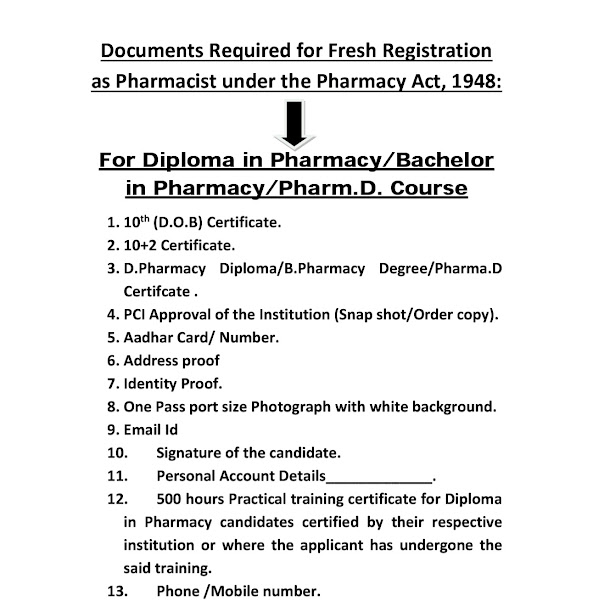
Documents Required for Fresh registration as pharmacist

          14. 3200/- for online registration charge.